
Department of Otolaryngology, Head and Neck Surgery
 |
Benign Lesions Department of Otolaryngology, Head and Neck Surgery |
| Normal |
Functional |
Infectious | Benign |
Malignant | Traumatic
| References |
| Home |
Patient History: This is a 47 year old patient who has had problems with his voice. He feels that there is constant hoarseness. He has no difficulty breathing.
Physical Findings: Reinke's space is immediately inferior to the mucosal squamous layer of the vocal cords and this is a space that allows the vocal folds to have the vibratory movement of the squamous layer. On this picture Reinke's space seems to have extra fluid in it and so there is slight bulging of the cords with swelling that can best be seen on the medial surface of both cords. When the cords vibrate, they frequently will not be in phase because of the fluid irregularities that occur.
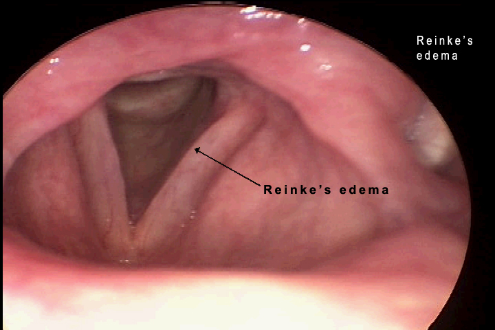
Comments: Reinke's edema is also known as polypoid degeneration and chronic edematous hypertrophy. It is seen in middle aged patients and up to 98 percent of them are smokers. Heavy smoking is a predisposing factor as well as working in a smoking environment, and a history of voice abuse. It is seen more frequently in females but this may only occur because it tends to lower the pitch of their voices and is most noticeable in a female voice. It is usually bilateral and chronic. The treatment is to stop smoking, decrease voice abuse and other environmental exposures. Surgery may be indicated if the edema becomes progressive.
Patient History: A 58 year old female who is here because of hoarseness. She has been treated for the last three years for airway problems including shortness of breath and severe hoarseness. This has increased severely over the last several months and her asthma medications were not helpful.
Physical Findings: The polyps are very extensive. They are long standing with a lot of fluid in the polyps on both sides. This is a continuation of Reinke's edema that has persisted for many years. These polyps can be seen to vibrate but are not in phase and cause hoarseness. The first photograph shows a large polyp on the left true vocal cord. This will cause breathiness in the voice because the true vocal cords cannot approximate each other and the wave will be disrupted on the left side. In the second photograph there is extensive disease in her airway on inspiration which was mistaken for asthma. These polyps can occur anywhere on the cords from a small area to the full length of the cord.
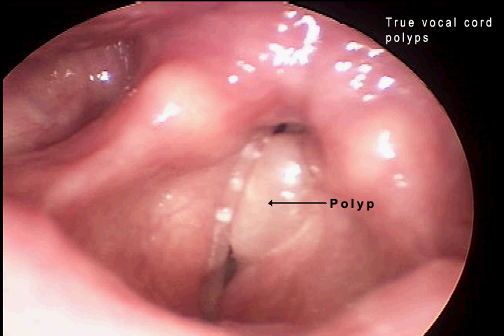
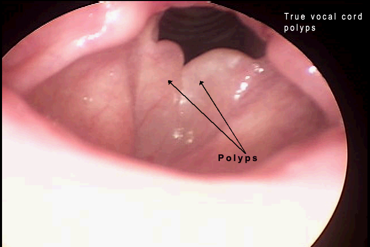
Comments: Most of the vocal cord polyps are benign lesions that usually occur because of voice abuse and are seen more frequently in women between the ages of twenty and forty. Ninety percent are unilateral, ten percent are bilateral. They are treated with surgical excision.
Patient History: This is a 19 year old female who has been hoarse for about a year. She has been a cheerleader at school and also participating in sports.
Note: No Picture or Video
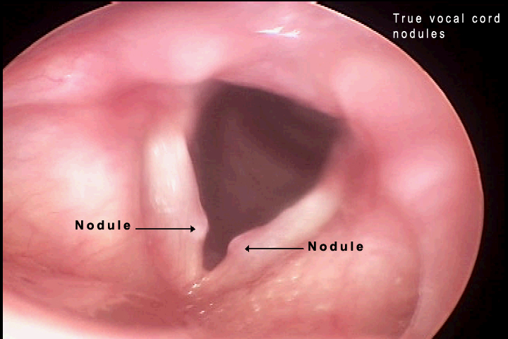
Patient History: This is a 36 year old female who has worked for several years at the casino within a loud environment and presented with increasing throat strain and breathiness. She has been hoarse for several years.
Physical Findings: The true vocal cord noules appear as single nodules on the medial surface of the vocal cords. They are not fluid filled and they do not change with the wave motion and frequently will have thickening on the most medial surface. This is noticeable in the second photograph where the nodules have been present for a long time. There is a figure of eight glottic closure which means that air is escaping both anteriorly and posteriorly but separated by the nodules. This gives a lot of breathiness and hoarseness.
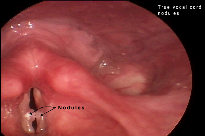
Comments: These are referred to as singers' nodules or screamers nodules. They arise in the junction between the anterior one-third and posterior two-thirds of the cords. They are flat, semi-translucent mucosal lesions. When they occur in children they are twice as common in boys, usually between the ages of five and fourteen, they are almost always bilateral and in children they will resolve spontaneously without surgery but with speech therapy. If the lesions are extremely large, they need to be removed surgically.
Patient History: This is a 42 year old female who is having problems with hoarseness for about three years. She had quit smoking but her voice did not improve.
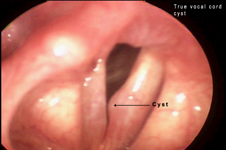
Patient History: A 48 year old school teacher who is finding that she is having more problems during the school year. She teaches music and also band.
Physical Findings: Notice the unilateral swelling on the medial surface of the vocal cords. There is no thickening. These are more fluid than the nodules and are covered by smooth mucosa.
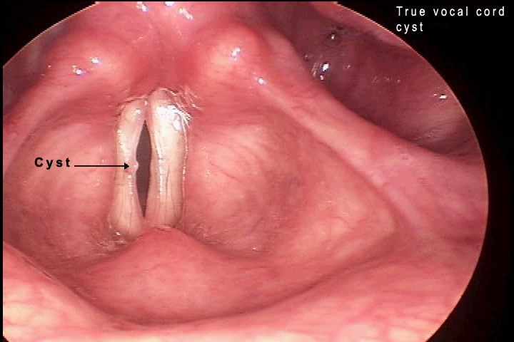
Comments: The cysts occur immediately beneath the squamous epithelium and are usually unilateral. Trauma to the vocal cords is considered the primary predisposing factor. These are removed surgically if they are interfering with the quality of the voice.
Patient History: This is a 22 year old female who underwent surgery several months ago and has a feeling of something in her throat since that time. Her voice is clear but she has frequent throat clearing and intermittent pain.
Physical Findings: The granuloma is a rounded mass erythematous in color, usually attached by a thinner stalk. Frequently it is superficial to the true vocal cord. It may affect the voice; however, the patient may have a normal voice with only a feeling of straining to talk.
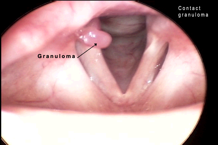
Comments: These are usually unilateral. One variety is caused because of intubation. These are surgically treated and tend to have a good resolution rate. The second kind of granuloma occurs with an ulcerated area on the medial surface of the arytenoid. There is frequently throat pain associated with these and it can be localized to one side of the neck. They may give referred pain to the ear and a feeling of a lump in the throat.There can be a high recurrance rate after surgical removal.
Patient History: This is a 55 year old gentleman who has been hoarse for six months. He quit smoking five months ago and had no improvement with antibiotics.
Physical Findings: The papillomas appear as small mulberry type lesions. They can attach anywhere in the larynx. Most frequently they start on the vocal cords. They decrease the wave motion and also the ability of the vocal cords to meet in the midline. In the photograph they can be seen in the anterior portion of the cords.
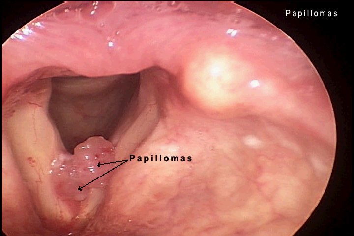
Comments: Papillomas are caused by the human papilloma virus (HPV), usually sub-types six and eleven. These occur frequently in children but can occur at any age. In adulthood they may grow very quickly and there are frequent recurrences. Surgery is indicated for their removal.
Patient History: This is a 60 year old female who has been experiencing some problems with her vocal range and came in for an evaluation of her larynx. The aryepiglottic cyst that was noted was giving her symptoms of a lump in the throat.
Physical Findings: The cysts can be seen on the left side of the aryepiglottic fold. It is more whitish in color than the surrounding mucosa and also is more elevated. It did not interfere with the speech nor does it change the ability of the vocal cords to move.
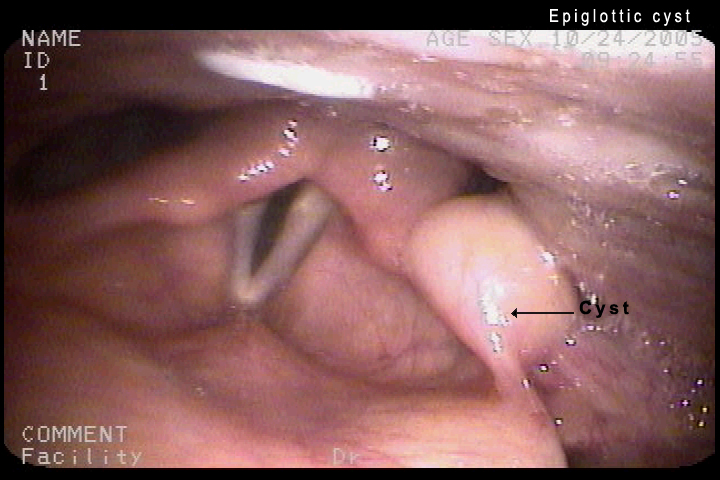
Comments: - Epiglottic cysts are benign cystic lesions that occur anywhere on the epiglottis, in the pre-epiglottic space or on the aryepiglottic folds. They most frequently occur on the lingual surface of the epiglottis. In general no medication is needed and surgery is only indicated for large cysts for the patients who have a globus sensation (feeling of a lump in the throat).
Patient History: This is a 45 year old female who smokes two packs per day. She works in the casino bar and has a lot of smoke exposure. She also has seasonal allergic rhinitis and a lot of post nasal drainage. She complains of cough, hoarseness and ear pain. She had no relief with antibiotic therapy.
Physical Findings: The physical findings show edema, erythema, some early changes of leukoplakia (white plaques), hyperemia of the entire area and loss of vocal cord wave formation.
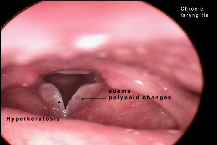
Comments: Chronic laryngitis frequently occurs in men. Ninety percent of patients are smokers. It can be caused by any type of inflammation, vocal abuse, reflux or allergies. Some times there are multiple causes. It is usually bilateral and there is thickening of the epithelium.
Patient History: This is a 35 year old female who has had a chronic cough, frequent throat clearing and varying degrees of hoarseness. She had proven acid reflux with laryngeal reflux on the pH probe at the region of the cricopharyngeus muscle, also known as the upper esophageal sphincter (UES).
Physical Findings: There is edema in both vocal cords and hypervascularity. Pachyderma, a condition of increasing mucosal tissue with swelling and hypertrophy in the area of the arytenoids, is seen in the space posterior to the vocal cords.
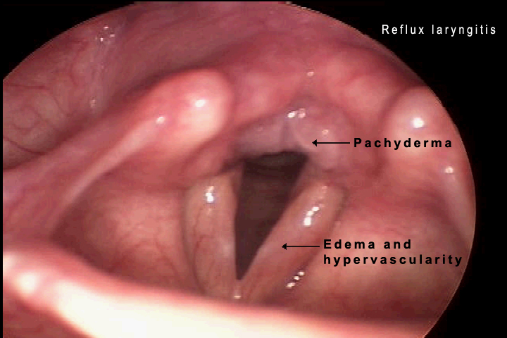
Comments: Laryngeal reflux is a cause of chronic hoarseness and one of the causes for chronic laryngitis. It is frequently seen in patients who have acid reflux up through the cricopharyngeus muscle (laryngopharyngeal reflux). It occurs most frequently in the upright position, during the daytime, and may lead to subglottic stenosis. This is in contrast to gastroesophageal reflux which usually occurs at night in the supine position.
Patient History: This is a 52 year old female who has had problems with hoarseness for several months and it is becoming worse.She has no problems with swallowing or breathing.
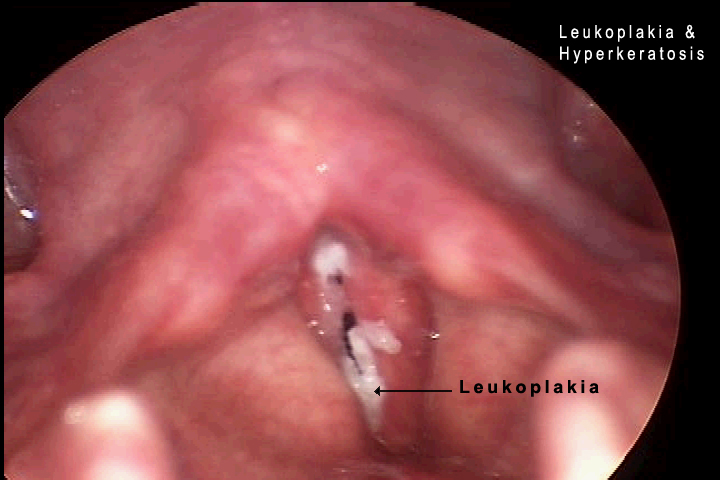
Patient History: This is a 55 year old gentleman who has chronic recurrent hoarseness. He has had multiple excisions of these lesions. His previous diagnoses have been hyperkeratosis.
Physical Findings: The patient has white plaques that are thickened in irregular parts of the vocal cords. This does not interfere with their abduction or adduction of the cords; however, there is a decrease in the wave formation and so the voice is not clear. On the second photograph there is extension to the medial edge, particularly on the left cord, so that there is markedly decreased wave formation with interference of speech.
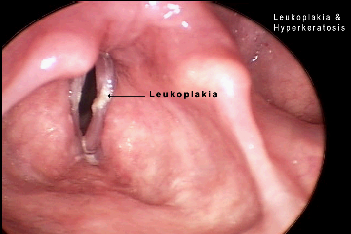
Comments: Hyperkeratosis is frequently a result of chronic laryngitis where there are permanent changes to the epithelium. It can be the development of extensive proliferation of the keratin. Leukoplakia is the name for the whitish plaques that can be seen on the vocal cords that are premalignant and often progress to squamous cell carcinoma.
Site administrator:
Barbara
Heywood MD.
Copyright © 2006
 All rights reserved.