
Department of Otolaryngology, Head and Neck Surgery
|
|
Normal Larynx Department of Otolaryngology, Head and Neck Surgery |
| Normal |
Functional |
Infectious | Benign |
Malignant | Traumatic
| References |
| Home |
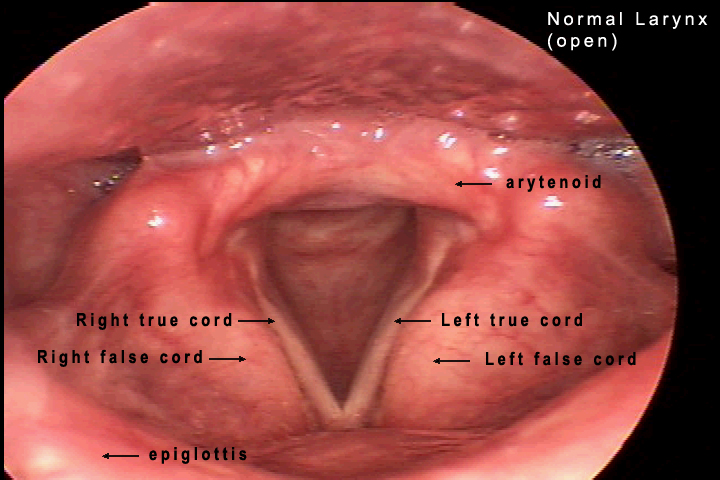
The following images show the larynx as it would appear if you were facing the patient looking down on the superior surface of the larynx. In the normal larynx, sound is produced by the two true vocal cords. In the first image, the cords are abducted or pulled open by the muscles of the larynx. You see a V shaped opening between the vocal cords through which you can see the tracheal cartilages below. The wide part of the V is posterior on the patient. The laryngeal structures are supported by cartilages which in turn are attached to the laryngeal muscles. The arytenoid cartilages are attached posteriorly to each vocal cord at their vocal processes and can be seen as prominent bulges lateral and posterior to the vocal cords. The posterior wall of the hypopharynx is in close proximity with the posterior part of the larynx. Between and beneath these two structures is the opening of the esophagus.
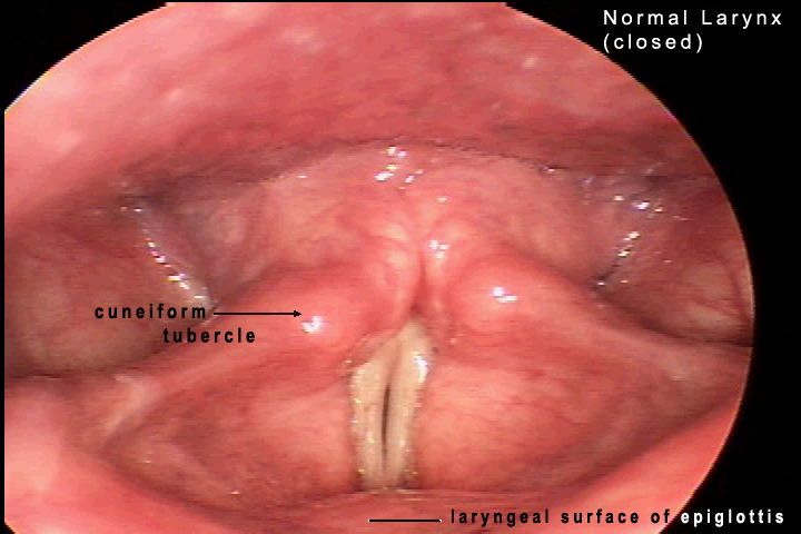
The second image shows the cords in an adducted position, pulled medially in approximation. Physiologically this approximation is necessary for protection of the airway during swallowing and to allow phonation. The outer mucosal covering of the vocal cords lies on a layer of collagenous tissue that allows the cords to vibrate in a characteristic way in response to air forced through the cords from the lungs. The vibration of the vocal cords causes a mucosal wave motion that produces the sound necessary for normal speech. On the normal video, observe the reflex abduction that occurs with inspiration and the adduction that accompanies phonation. You can also observe the mucosal wave which has been slowed and made visible by this stroboscopic video. Click on either image to launch the video.
Site administrator:
Barbara
Heywood MD.
Copyright © 2006
 All rights reserved.