Department of Otolaryngology, Head and Neck Surgery
 |
Otology Department of Otolaryngology, Head and Neck Surgery |
| Home | Unit One | Unit Two | Unit Three | Unit Four | Unit Five | Unit Six | Unit Seven | Unit Eight | References |
Examples of audiograms and typmpanograms
Estimated time to complete: X minutes. Please complete the entire unit in one access period.
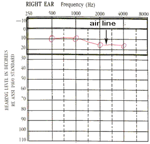
 Normal AudiogramA hearing test or audiogram measures the ability of the patient to hear sounds by air conduction and by bone conduction. The air conduction test is performed first and shown in this example. It measures hearing through the tympanic membrane and the ossicles. A TM perforation, middle ear fluid or an interruption in the bones would decrease the patient's air conduction level. This is the test frequently done in school or work screenings called an air screen. The bone conduction test measures the level of hearing through the skull and directly to the cochlea, bypassing the middle ear. A bone conduction device is placed over the mastoid bone behind the ear and sounds are presented. Normal hearing will be represented on an audiogram in the range of -10 to 20 decibels, with the air and bone levels equal. If the air line is normal, bone conduction is not usually tested. |
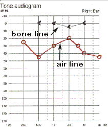
 Audiogram with conductive hearing lossIf the air line (red circles) is down and the bone line (black arrows) is normal, this is a conductive loss because the nerve is normal but the transmission through the TM and middle ear is decreased. The air level is never better than the bone unless the patient is tired or unable to cooperate. The conductive loss can be verified with tuning forks: The 256Hz and 512Hz forks will show that the bone hearing is better than air utilizing the Rinne test. |
 Audiogram with Mixed LossThe audiogram with a mixed loss shows a nerve loss when tested behind the ear and a larger loss when tested with the earphones. This occurs in a patient who has nerve damage and also a conductive component secondary to middle ear or external ear disease. The nerve line (black brackets) is at 55-60dB and the air line (red circles) averages 80dB. This 20dB difference is the conductive loss due to middle or external ear abnormalities. Examples of this are canal obstruction, tympanic membrane perforation, and serous otitis in a patient with underlying nerve loss. |
 Normal TympanogramTympanometry is a method of evaluating the ear drum mobility and condition of the middle ear. A probe is placed into the ear canal and air pressure varying from -400 to +200 is applied. As the pressure is changed in the external canal, the compliance (inverse of stiffness) of the tympanic membrane is simultaneously measured and recorded. This recording represents the compliance of a TM that is normal (Type A). The peak compliance is at 0. The TM is maximally compliant when the outer canal pressure is equal to the middle ear pressure and there is no middle ear pathology. It follows that the middle ear pressure is also 0. Tympanometry utilizes smaller pressures than pneumo-otoscopy and is able to measure minimal changes. The volume of the exteral ear canal is also measured, and in this case is 1.4 ml, normal for an adult ear with an intact TM. Adult canals are usually < 2.5ml, the external canals of children are usually less than 1.0 ml. These volumes vary with the age of the patient and the size of the canal. Volume also varies with the size of the ear probe. A small ear probe inserted deeply in the canal makes the volume much smaller. |
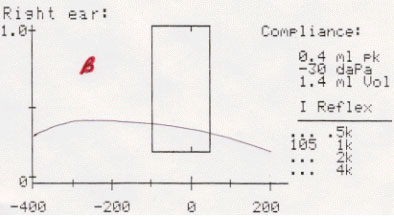
 Flat TympanogramThis recording illustrates a "flat" tympanogram, designated type B. There is little change in compliance as external canal pressure is varied. This implies a relatively immobile TM. This commonly occurs in four cases: 1) The middle ear space is filled with fluid (serous otitis media) - in this case, the volume will be normal.2) The tympanic membrane is severely retracted and draped over the ossicles obliterating the middle ear space so that it can no longer vibrate (adhesive otitis media) - in this case the volume will be normal or slightly higher. 3) The tympanic membrane has a perforation, either a healed opening or maintained by a myringotomy tube - in this case the volume will be very large, since the tympanogram is measuring the additional volume of the middle ear and the mastoid. 4) The ear probe is incorrectly placed against the ear canal, blocking it - in this case, the graph will have a very small volume. |
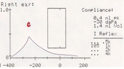
 Negative Pressure TympanogramThis tympanogram illustrates decreased compliance and and the maximum compliance occurs at a negative pressure. It is designated type C. The patient has negative middle ear pressure reducing the mobility of the TM. Since air is actively absorbed from the middle ear through the mucosa, an inability to open the eustachian tube results in a negative pressure. This finding is seen in patients who have eustacean tube dysfunction for a variety of reasons including upper respiratory infection, allergy or radiation therapy to the head. |
| Home | Unit One | Unit Two | Unit Three | Unit Four | Unit Five | Unit Six | Unit Seven | Unit Eight | References |
Site administrator:
Barbara
Heywood MD.
Copyright © 2014
 All rights reserved.