Department of Otolaryngology, Head and Neck Surgery
 |
Otology Department of Otolaryngology, Head and Neck Surgery |
| Home | Unit One | Unit Two | Unit Three | Unit Four | Unit Five | Unit Six | Unit Seven | Unit Eight | References |
History:
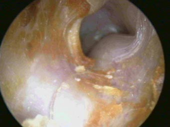
This is a 24 year old female who was seen in the office for pulsatile tinnitus. She had no complaints of hearing loss, ear pain or ear infections.
Examination:
The inferior portion of the tympanic membrane appears erythematous. There is a lesion medial to the tympanic membrane. This proved to be a glomus tympanicum tumor.
 |
^ Click on the arrow to view the video |
Information: Paragangliomas of the ear are benign, slow-growing vascular lesions that arise from a glomus body in the temporal bone or jugular bulb. Formerly known as glomus tumors, these are the most common benign tumors to arise in the temporal bone. These tumors may rarely secrete catecholamines and other vasoactive substances such as noradrenaline, adrenaline, dopamine and serotonin. They have a 10% rate of malignant transformation. Site of origin is determined by examining the cellular composition of the tumor. The tumors are similar in cellular composition to the tissue from which they originate, and are composed largely of precapillary or capillary vessels. From the site of origin, the tumor grows and infiltrates nearby structures. Commonly it compresses the sigmoid sinus and/or jugular bulb or it protrudes into the bulb. On tissue examination, the tumor appears with a rich supply of vascular spaces lined by epithelial cells. The cells are uniform in size and appearance. Those that invade surrounding tissue are pleomorphic, invading bone, making it soft and hemorrhagic. At surgery, the tumor is hemorrhagic because the vascular spaces have no contractile elements.
Signs and symptoms include pulsatiletinnitus, secondary infection, bleeding, discharge, hearing loss,otalgia, and enlargement behind the tympanic membrane, invading and destroying the incus.
Brown’s sign is evident during pneumo-otoscopy demonstrating blanching of the mass with the application of positive pressure. Diagnosis is confirmed by radiological studies. Early tumors appear as a reddish,pulsatile swelling in the middle ear. The tumor may erode the TM, forming a smooth, dark red, polypoid mass that bleeds easily with manipulation. Females are three times as likely than males to have a paraganglioma with most tumors occurring in middle age Caucasians.
Glomus tympanicum - A paraganglioma arising in the middle ear space. A red-blue retro tympanic mass may be seen in 50% of the tumors. Glomus jugulare - is a paraganglioma arising from the jugular bulb at the jugular foramen. These tumors tend to to be less symptomatic and achieve a larger size before discovery. They frequently invade the base of the skull and temporal bone and often present as a lesion in the hypotympanum. It is impossible to differentiate a glomus tympanicum from a glomus juglulare invading the hypotympanum without radiographic imaging.
Glomus tympanicum. This is an 82 year old female with left sided pulsatile tinnitus of one year’s duration. Note the erythema and pulsation of the glomus tympanicum tumor, which fills the middle ear space.
 |
OsteomaOsteomas are benign bony tumors. They occur at the bony-cartilaginous junction of the tympanomastoid suture line. Removal is needed only if they become obstructive, which is rare. |
| Home | Unit One | Unit Two | Unit Three | Unit Four | Unit Five | Unit Six | Unit Seven | Unit Eight | References |
Site administrator:
Barbara
Heywood MD.
Copyright © 2014
 All rights reserved.